ƽ

STEMdiff™ Cerebral Organoid Maturation Kit

Culture medium kit for extended maturation of human cerebral organoids

STEMdiff™ Cerebral Organoid Maturation Kit

Culture medium kit for extended maturation of human cerebral organoids

Catalog #
(Select a product)
Culture medium kit for extended maturation of human cerebral organoids
Request Pricing Request Pricing

Product Advantages

  • Generate unpatterned organoids capable of spontaneous differentiation to produce multiple brain regions within the same organoid
  • Culture under flexible conditions with either matrix droplet embedding or liquid matrix
  • Enjoy increased efficiency of organoid formation with a formulation based on a popular published protocol
  • Generate new or modified organoid models with this highly compatible platform

What's Included

  • STEMdiff™ Cerebral Organoid Basal Medium 2, 250 mL
  • STEMdiff™ Cerebral Organoid Supplement E, 4.5 mL
Products for Your Protocol
To see all required products for your protocol, please consult the Protocols and Documentation.

What Our Scientist Says

Human brain development is complex, so to observe it in a dish is a huge scientific breakthrough. We've tried to simplify that process and make it more accessible to you, regardless of how much stem cell experience you have.

Leon ChewScientist
Leon Chew, Scientist

Overview

Culture pluripotent stem cell (PSC)-derived neural organoids for extended periods (> 40 days) with the STEMdiff™ Cerebral Organoid Maturation Kit.

For your convenience, the maturation kit is fully compatible with the STEMdiff™ Cerebral Organoid Kit, which can be used to generate neural organoids under defined and serum-free conditions.

For more information on protocols for organoid culture with STEMdiff™ Cerebral Organoid Maturation Kit, please explore the Product Information Sheet (PIS) and Educational Materials.
Subtype
Specialized Media
Cell Type
Neural Cells, PSC-Derived, Neural Stem and Progenitor Cells, Pluripotent Stem Cells
Species
Human
Application
Cell Culture, Characterization, Differentiation, Functional Assay, Immunofluorescence, Organoid Culture, Phenotyping, Spheroid Culture
Brand
STEMdiff
Area of Interest
Disease Modeling, Neuroscience, Stem Cell Biology
Formulation Category
Serum-Free

Data Figures

Cerebral Organoids Contain Multiple Layered Regions That Recapitulate the Cortical Lamination Process Observed During In Vivo Human Brain Development

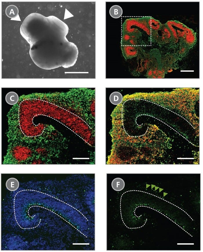
Figure 1. Cerebral Organoids Contain Multiple Layered Regions That Recapitulate the Cortical Lamination Process Observed During In Vivo Human Brain Development

(A) A representative phase-contrast image of a whole cerebral organoid at Day 40 generated using the STEMdiff™ Cerebral Organoid Kit. Cerebral organoids at this stage are made up of phase-dark structures that may be surrounded by regions of thinner, more translucent structures that display layering (arrowheads). (B) Immunohistological analysis on cryosections of cerebral organoids reveals cortical regions within the organoid labeled by the apical progenitor marker PAX6 (red) and neuronal marker β-tubulin III+ (TUJ-1) (green). (C-F) Inset of boxed region from (B). (C) PAX6+ apical progenitors (red, enclosed by dotted line) are localized to a ventricular zone-like region. β-tubulin III+ neurons (green) are adjacent to the ventricular zone. (D) CTIP2, a marker of the developing cortical plate, co-localizes with β-tubulin III+ neurons in a cortical plate-like region. Organization of the layers recapitulates early corticogenesis observed during human brain development. (E) Proliferating progenitor cells labeled by Ki-67 (green) localize along the ventricle, nuclei are counterstained with DAPI (blue). (F) An additional population of Ki-67+ cells is found in an outer subventricular zone-like region (arrowheads). Scale bar = (A) 1 mm, (B) 1 mm and (C-F) 200 µm.

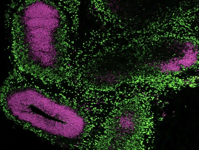
Immunocytochemistry image of a cerebral organoid cultured in mTeSR™ Plus and directed to cerebral organoids using the STEMdiff™ Cerebral Organoid Kit.

Figure 2. Cerebral Organoids Can Be Generated from hPSCs Maintained in mTeSR™ Plus

Human ES (H9) cells were cultured with mTeSR™ Plus and directed to cerebral organoids using the STEMdiff™ Cerebral Organoid Kit. Image shows apical progenitor marker SOX2 (magenta) and neuronal marker TBR1 (green).

Cryosectioned Cerebral Organoids Show Stratification of Cortical Plate Neurons and Progenitor Zones

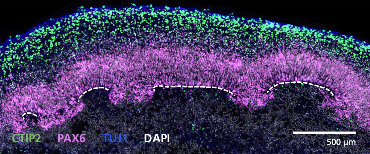
Figure 3. Cryosectioned Cerebral Organoids Show Stratification of Cortical Plate Neurons and Progenitor Zones

Cerebral organoids were generated using the STEMdiff™ Cerebral Organoid Kit. A 16-μm-thick section of a Day 40 cerebral organoid was stained for CTIP2 (green), PAX6 (magenta), βIII-tubulin/TUJ1 (blue), and DAPI (gray). Cortical regions are defined by progenitor cells (PAX6+) that are radially organized around a pseudo-ventricle (dashed line). These progenitors give rise to cortical plate neurons indicated by CTIP2 and TUJ1 expression. For a detailed cryogenic tissue processing and immunofluorescence protocol, please see the Methods Library.

Zones of Active Proliferation in Cerebral Organoids Are Preserved Following the Protocol for Tissue Processing

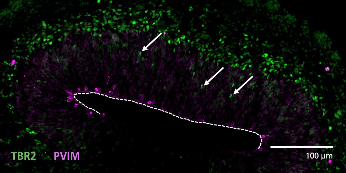
Figure 4. Zones of Active Proliferation in Cerebral Organoids Are Preserved Following the Protocol for Tissue Processing

Organoid tissue was processed for immunofluorescence and stained for TBR2 (intermediate precursors, green) and phosphorylated vimentin (PVIM, dividing cells, magenta). Cells actively divide at the apical border of cortical regions along the border of the pseudo-ventricle (dashed line). A population of these dividing cells will express TBR2 and then migrate (arrows) from the progenitor zone to form a layer of intermediate progenitors. For a detailed cryogenic tissue processing and immunofluorescence protocol, please see the Methods Library.

Immunofluorescence from Cryosectioned Cerebral Organoids Indicates Preserved Organization of Cortical Neurons

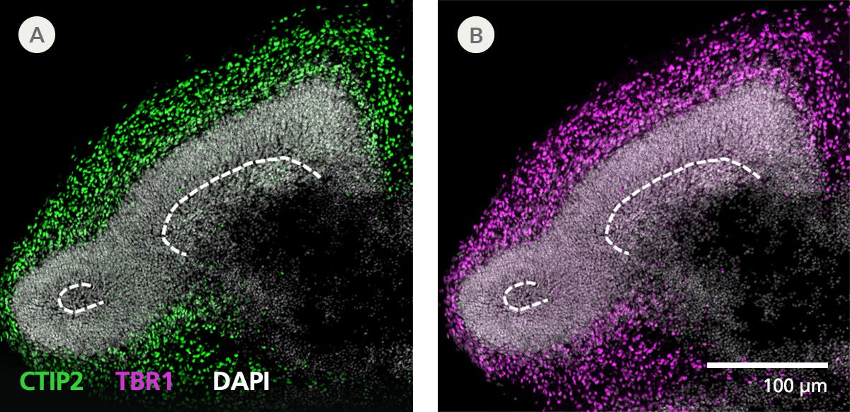
Figure 5. Immunofluorescence from Cryosectioned Cerebral Organoids Indicates Preserved Organization of Cortical Neurons

Organoid tissue was processed for immunofluorescence and stained for CTIP2 (green), TBR1 (layer 5/6 cortical neurons, magenta), and DAPI (white). Deep layer neuronal markers CTIP2 and TBR1 are expressed in cells around presumptive progenitor zones (dashed line) toward the outside or apical surface of organoids. For a detailed cryogenic tissue processing and immunofluorescence protocol, please see the Methods Library.

Cryogenic Tissue Processing and Immunofluorescence Captures Arrangement of Neural Progenitors Around Pseudo-Ventricles in Cerebral Organoid

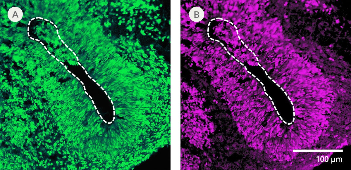
Figure 6. Cryogenic Tissue Processing and Immunofluorescence Captures Arrangement of Neural Progenitors Around Pseudo-Ventricles in Cerebral Organoids

Organoid tissue was processed for immunofluorescence and stained for (A) FOXG1 (forebrain cells, green) or (B) SOX2 (neural progenitors, magenta). Organoids derived from STEMdiff™ Cerebral Organoid Kit generate forebrain-type tissue as indicated by FOXG1 expression. Neural progenitors expressing SOX2 are radially arranged around a pseudo-ventricle area (dashed line). For a detailed cryogenic tissue processing and immunofluorescence protocol, please see the Methods Library.

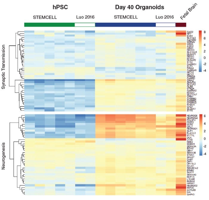
Neural Organoids Generated with STEMdiff™ Cerebral Organoid Kit Express Expected Key Markers

Figure 7. Neural Organoids Generated with STEMdiff™ Cerebral Organoid Kit Express Expected Key Markers

Heatmap of expression levels for genes associated with synaptic transmission function and neurogenesis in Day 40 organoids. These data show that gene expression of cerebral organoids generated from the STEMdiff™ Cerebral Organoid Kit are similar to published results (C Luo et al. Cell Rep, 2016).

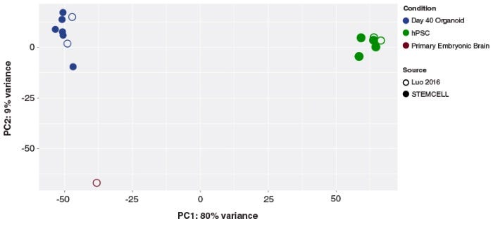
Cerebral Organoids Generated with the STEMdiff™ Cerebral Organoid Kit Are Transcriptionally Similar to Those from Published Protocols

Figure 8. Cerebral Organoids Generated with the STEMdiff™ Cerebral Organoid Kit Are Transcriptionally Similar to Those from Published Protocols

Principal component analysis of hPSC and cerebral organoid transcriptomes. Cerebral organoids generated using the STEMdiff™ Cerebral Organoid Kit (filled blue circles) cluster together, and cluster with previously published (C Luo et al. Cell Rep, 2016) cerebral organoids (open blue circles). The first principal component accounts for the majority of variance seen (PC1; 80%) and distinguishes the cerebral organoid samples from the hPSCs (green circles). The second principal component accounts for only 9% of the variation, and highlights the modest expression differences between cultured organoids and primary embryonic fetal brain samples (19 post-conceptional weeks, brown circles).

Time-lapse images and heatmap showing calcium signals spreading across a Day 91 cerebral organoid, indicating synchronized neuronal activity.

Figure 9. STEMdiff™ Cerebral Organoids Exhibit Synchronous Calcium Activity

Cerebral organoids were generated using the STEMdiff™ Cerebral Organoid Kit and matured with the STEMdiff™ Cerebral Organoid Maturation Kit. On Day 91, organoids were imaged using the with MetaXpress® High-Content Image Acquisition and Analysis Software. (A) Time-lapse images of a representative region within a cerebral organoid. Arrows indicate areas of elevated calcium signal intensity. (B) Corresponding heatmap representation of calcium intensity from the regions shown in (A) over time. Signal propagation from the initial peak to surrounding areas suggests the presence of a functional neuronal network.

Protocols and Documentation

Find supporting information and directions for use in the Product Information Sheet or explore additional protocols below.

Document Type
Product Name
Catalog #
Lot #
Language
Document Type
Product Name
Catalog #
08571
Lot #
All
Language
English
Document Type
Product Name
Catalog #
08571
Lot #
All
Language
English
Document Type
Product Name
Catalog #
08571
Lot #
All
Language
English
Document Type
Product Name
Catalog #
08571
Lot #
All
Language
English
Document Type
Product Name
Catalog #
08571
Lot #
All
Language
English
Document Type
Product Name
Catalog #
08571
Lot #
All
Language
English
Document Type
Product Name
Catalog #
08571
Lot #
All
Language
English

Applications

This product is designed for use in the following research area(s) as part of the highlighted workflow stage(s). Explore these workflows to learn more about the other products we offer to support each research area.

Resources and Publications

Educational Materials (41)

Brochure
Brochure
Brochure
Scientific Poster
Scientific Poster
Scientific Poster
Scientific Poster
Scientific Poster

Publications (12)

A human cerebral organoid model of West Nile virus encephalitis shows innate immunocompetency J. F. Steffen et al. Nature Communications 2026 Mar

Abstract

West Nile virus (WNV), an arbovirus of emerging global interest, can cause neuroinvasive disease in humans. Currently, no protective vaccine or specific treatment is available for human WNV encephalitis. The virus induces neuronal cell death, while astrocytes and microglia cells are suspected to contribute to WNV pathology. Hence, understanding their role is crucial for future treatment approaches. In this study, we establish a WNV encephalitis model using human cerebral organoids, generated with male iPSCs. Infection results in heterogeneous kinetics with an early strong replication potentially leading to viral clearance, while a late peak was associated with more long-term infection. Viral foci are seen in cortical-like areas, rich in neurons and astrocytes, however void of microglia. Pro-inflammatory cytokines (IL-6, TNF-α, IL-18), chemokines (CXCL10, CCL17, CX3CL1, CCL2) and biomarkers (IL-1RA, sTREM-1, sRAGE, BDNF) are increasingly released. Conclusively, human cerebral organoids make suitable WNV encephalitis models with valuable properties to study acute and long-term infection. West Nile virus (WNV) can cause neuroinvasive disease. Here the authors develop a human cerebral organoid model for WNV infection and find heterogeneous viral kinetics with viral foci in neuron- and astrocyte-rich areas devoid of microglia, as well as increased release of cytokines and other biomarkers.
Early-life exposure to polypropylene nanoplastics induces neurodevelopmental toxicity in mice and human iPSC-derived cerebral organoids F. Huang et al. Journal of Nanobiotechnology 2025 Jul

Abstract

Nanoplastics (NPs) are emerging environmental pollutants that pose growing concerns due to their potential health risks. However, the effects of inhaled NP exposure during pregnancy on fetal brain development remain poorly understood. In this study, we investigated the impact of maternal exposure to polypropylene nanoplastics (PP-NPs) on fetal brain development and neurobehavioral outcomes in a mouse model and further explored its mechanism in human cerebral organoids. Maternal exposure to PP-NPs significantly impaired neuronal differentiation and proliferation in the fetal cortex. Neurobehavioral assessments revealed significant deficits in offspring following maternal exposure, including impaired spatial memory, reduced motor coordination, and heightened anxiety-like behavior. Furthermore, human brain organoids exposed to PP-NPs exhibited reduced growth and neuronal differentiation, with significant downregulation of key neuronal markers such as TUJ1, MAP2, and PAX6. Transcriptomic analysis identified alterations in gene expression, particularly in neuroactive ligand-receptor interaction pathway. Molecular docking and fluorescence co-localization analysis further suggested CYSLTR1 and PTH1R as key molecular targets of PP-NPs. These findings provide novel insights into the toxicological effects of NPs on the developing brain and emphasize the need for preventive measures to protect fetal neurodevelopment during pregnancy. The online version contains supplementary material available at 10.1186/s12951-025-03561-1.
RNA Sequencing Reveals a Strong Predominance of THRA Splicing Isoform 2 in the Developing and Adult Human Brain E. Graceffo et al. International Journal of Molecular Sciences 2024 Sep

Abstract

Thyroid hormone receptor alpha (THRα) is a nuclear hormone receptor that binds triiodothyronine (T3) and acts as an important transcription factor in development, metabolism, and reproduction. In mammals, THRα has two major splicing isoforms, THRα1 and THRα2. The better-characterized isoform, THRα1, is a transcriptional stimulator of genes involved in cell metabolism and growth. The less-well-characterized isoform, THRα2, lacks the ligand-binding domain (LBD) and is thought to act as an inhibitor of THRα1 activity. The ratio of THRα1 to THRα2 splicing isoforms is therefore critical for transcriptional regulation in different tissues and during development. However, the expression patterns of both isoforms have not been studied in healthy human tissues or in the developing brain. Given the lack of commercially available isoform-specific antibodies, we addressed this question by analyzing four bulk RNA-sequencing datasets and two scRNA-sequencing datasets to determine the RNA expression levels of human THRA1 and THRA2 transcripts in healthy adult tissues and in the developing brain. We demonstrate how 10X Chromium scRNA-seq datasets can be used to perform splicing-sensitive analyses of isoforms that differ at the 3′-end. In all datasets, we found a strong predominance of THRA2 transcripts at all examined stages of human brain development and in the central nervous system of healthy human adults.